Sample reads

See what comes back at 2 AM.

Seven common overnight studies, with the findings and impression you would see in your system. Illustrative cases, fictional patients.

  • HIPAA + BAA
  • NCQA-certified CVO
  • Joint Commission standards
  • Finals read in the US
  • Live in 30 days

Case library

Seven reads, start to finish.

Stroke, chest, abdomen, head trauma and spine: the studies that fill a night.

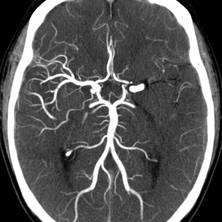
Illustrative CT angiogram of the head showing a left M1 middle cerebral artery occlusion

CTA head · stroke protocol

Stroke CTA

Findings
Abrupt cut-off of the proximal left M1 MCA. Reduced filling of the left MCA branches. No hemorrhage.
Impression
Acute left M1 large vessel occlusion.
Critical result
Called to the stroke team, read-back confirmed.
Illustrative · fictional patient
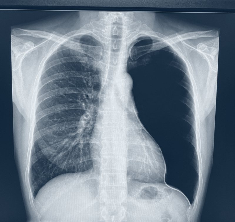
Illustrative upright chest X-ray showing a large left pneumothorax

Chest X-ray · PA upright

Pneumothorax

Findings
Large left pneumothorax with a visible pleural line. Mild mediastinal shift.
Impression
Large left pneumothorax.
Critical result
Called to the ED physician, read-back confirmed.
Illustrative · fictional patient
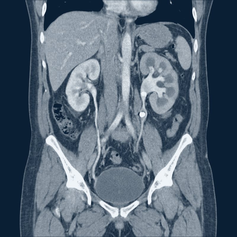
Illustrative coronal CT of the kidneys and ureters showing an obstructing left ureteral stone

CT KUB · coronal

Kidney stone

Findings
6 mm stone in the left proximal ureter. Mild left hydronephrosis.
Impression
Obstructing left ureteral calculus.
Illustrative · fictional patient
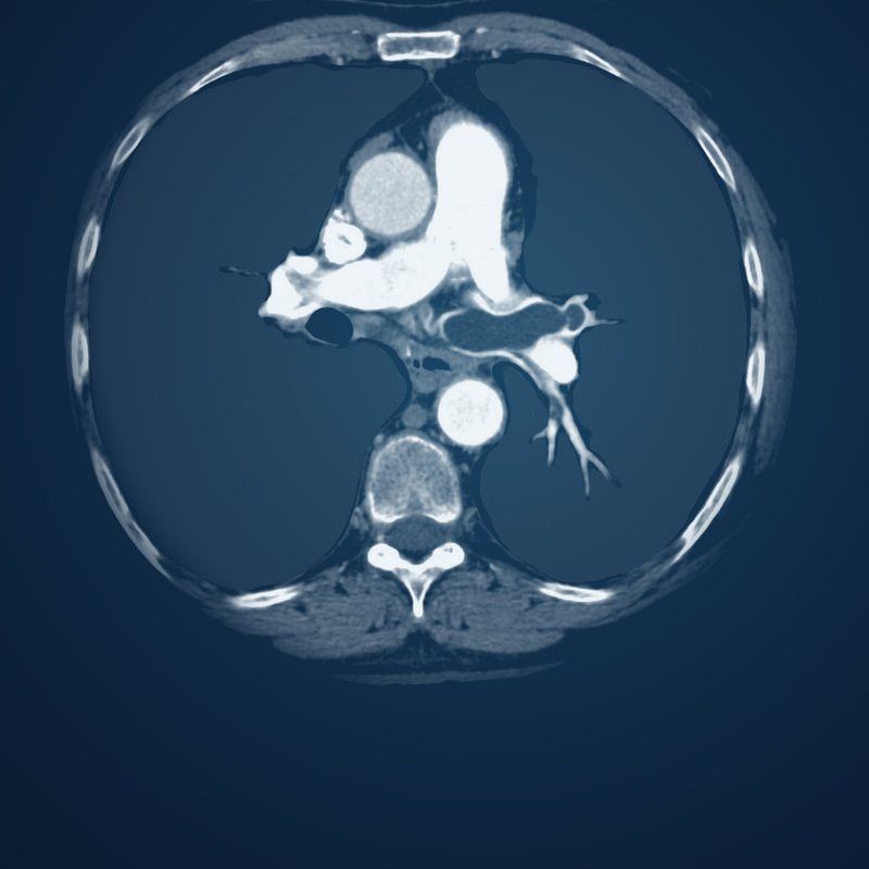
Illustrative CT pulmonary angiogram showing a filling defect in the left pulmonary artery

CTA chest · PE protocol

Pulmonary embolism

Findings
Filling defect in the left main pulmonary artery, extending into the left lower lobe branches. RV/LV ratio 1.1.
Impression
Acute pulmonary embolism.
Critical result
Called to the ED physician, read-back confirmed.
Illustrative · fictional patient
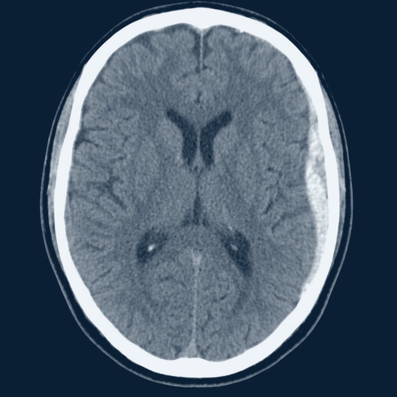
Illustrative non-contrast head CT showing an acute left subdural hematoma

CT head without contrast

Head trauma · subdural

Findings
Acute left convexity subdural hematoma, about 1 cm thick. Mild midline shift.
Impression
Acute left subdural hematoma with mass effect.
Critical result
Called to the ED physician, read-back confirmed.
Illustrative · fictional patient
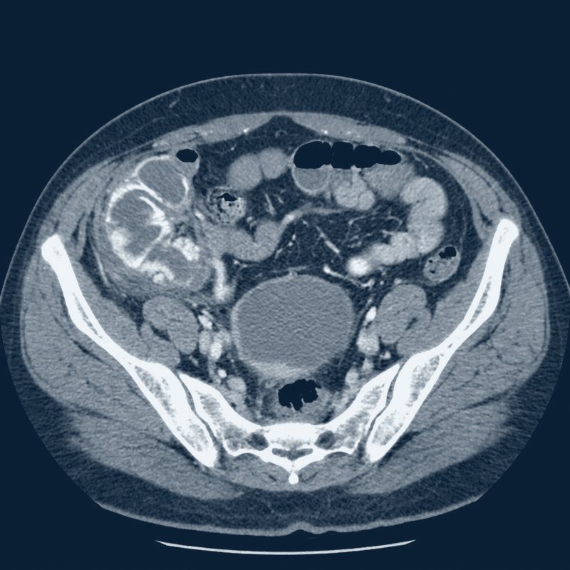
Illustrative contrast CT of the abdomen and pelvis showing acute appendicitis

CT abdomen / pelvis with contrast

Appendicitis

Findings
Dilated, thick-walled appendix with periappendiceal fat stranding. No abscess or free air.
Impression
Acute appendicitis.
Critical result
Surgery paged.
Illustrative · fictional patient
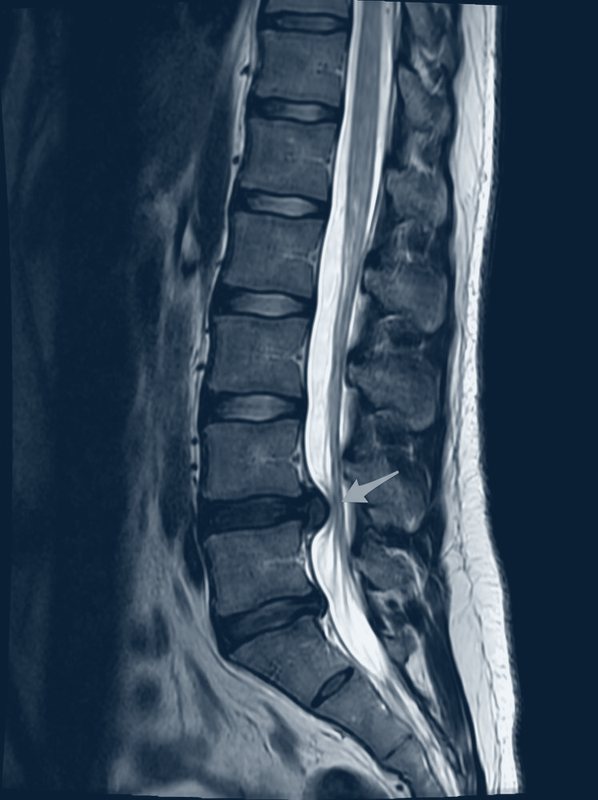
Illustrative sagittal T2 MRI of the lumbar spine showing an L4–L5 disc extrusion

MRI lumbar spine · T2 sagittal

Disc herniation

Findings
L4–L5 disc extrusion narrowing the left lateral recess.
Impression
L4–5 disc extrusion with left L5 nerve root impingement.
Illustrative · fictional patient

The report

What your team gets.

In every report

  • A signed final by a board-certified radiologist
  • Your templates and your critical list
  • Findings and a clear impression
  • Critical results called and documented with read-back

About these cases

Every case on this page is illustrative. The patients are fictional and no real patient information is shown. The images show the kinds of studies we read every night.

Watch a stroke get read

Related guides

Want to see reads in your format?

We’ll walk through your templates and critical list on your coverage plan call.

Get my free coverage plan